A
novel, non-contact ultrasound device is detailed for recording and analyzing 3D
fast eye movements (saccades) and smooth pursuit eye movements. Saccades are
studied to gain a better understanding of the human oculomotor plant and
neuromuscular systems. Abnormal saccades can be indicators of both neurological disorders and mild traumatic brain injury (MTBI). Limitations in existing
saccade measurement devices prevent them from being used to measure saccades
immediately after a possible MTBI event or easily outside of the clinical
environment.

Data obtained indicated no cytotoxic
response in control cells, as the viability of cells without treatment with AF
standard or methanolic extracts of AF extracts [negative control] using
methanol as the reconstituting solvent, was 99.9% after 24 hrs. of incubation.
However, cell viability significantly (p<0.001) decreased upon exposure to AF extracts especially for poultry feed. This was influenced by both the dose
and duration of exposure, which was much more pronounced when the cells were
exposed to AFB1 standard than for all the AF extracts tested. This implies that
these feeds on exposure to AF can greatly influence animal health with respect
to both the contamination dose and exposure time.

We propose that bioengineered cranial bones with multiple
intelligent functions, including site specific Tran’s meningeal drug delivery
and neurotoxin drainage with EEG feedback, can provide effective treatment of these
brain disorders by drug combinations that act on both synapses and genes with
concomitant selective drainage of harmful extracellular molecules. After examining and summarizing the rationale and feasibility of this proposal, we
suggest novel methods for extending the functions of the involved components
including synergies with existing devices and we highlight relevant pre clinical
results, discussing medical prospects of this novel neuro therapeutic approach.
Finally, we discuss key engineering, scientific, clinical and ethical
challenges to introducing bio engineered cranial bones with multiple intelligent
functions to the clinic within a decade.

Histological criteria for the
diagnosis of IDC-P include solid; dense cribriform (>50% cellularity of the
lumen); trabecular/micropapillary; and loose cribriform intraductal proliferation
of malignant cells. The latter two growth patterns share much similarity with HGPIN. In these instances, additional diagnostic criteria, such as marked
nuclear pleomorphism (nuclear enlargement > 6x normal nuclei), and nonfocal
comedonecrosis (> 1 duct showing comedonecrosis) are criteria needed to
differentiate it from HGPIN.

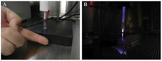
The advantage of cold plasma therapy
over conventional thermal plasma treatments, arc coagulators and desiccators,
is that it allows for more precise application and therefore more controllable
effects on the tissue. Additionally, cold plasma treatment showed stimulatory effects on wound healing and tissue regeneration. Experiments show that cold
atmospheric plasma treatment allows for efficient, non-contact, painless, and
antiseptic effects without damaging healthy tissue. As a result of the better
understanding of complex plasma phenomena and the development of new plasma
sources in the past few years, plasma medicine has developed into an innovative
and promising field of research.
Current methods for the assessment
of the outcome after anterior knee pain or lateral patellar instability
treatment have several limitations, for example their subjectivity. Therefore,
new technologies are needed to objectively evaluate the outcomes of treatments
for patellofemoral disorders.
Kinematic and kinetic analyses during dynamic
activities under realistic loading conditions that trigger or aggravate the
symptoms can: evaluate the patellofemoral patient in an objective way before
surgery; analyse the defense mechanisms the patient develops in order to reduce pain and/or instability; improve our knowledge of the aetiopathogeny and
therefore of a suitable treatment for patellofemoral disorders; and objectively
evaluate the result of the treatment. However, the kinetic and kinematic
analyses are not diagnostic tools.


